

The microscope worksheet is an essential educational resource, helping students identify and understand microscope components, functions, and practical uses. Suitable for multiple grade levels with answer keys provided.
1.1 Overview of the Microscope Worksheet
The microscope worksheet provides a comprehensive guide for students to learn about microscope components, their functions, and proper usage. It includes labeling exercises, diagrams, and practical examples. Worksheets are designed for various grade levels, offering interactive learning experiences. Answer keys are included for self-assessment, ensuring understanding of key concepts. This resource is ideal for biology and science education, promoting hands-on learning and visual engagement.
1.2 Importance of Microscope Worksheets in Education
Microscope worksheets are vital educational tools, enhancing students’ understanding of microscopy concepts. They provide interactive learning, improving retention and practical skills. Worksheets cater to diverse learning styles, offering clear diagrams and exercises. Answer keys enable self-assessment, while labeling and functional analysis deepen knowledge. These resources prepare students for lab work, fostering confidence in handling microscopes and interpreting results effectively in science education.
Labeling the Parts of the Microscope
Labeling microscope parts helps students identify and understand their roles, making complex concepts accessible through interactive and hands-on learning experiences with clear diagrams and exercises.
2.1 Identifying the Key Components
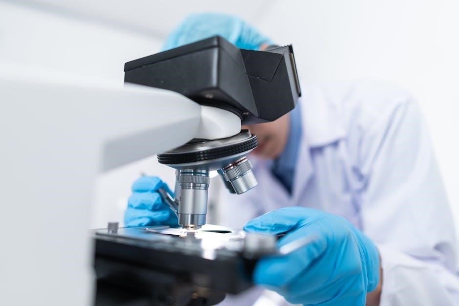
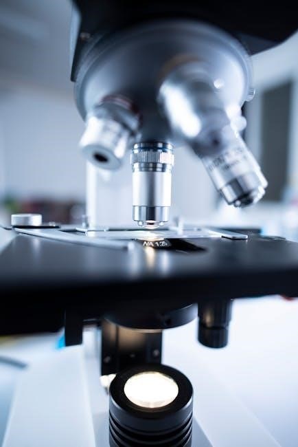
Identifying key microscope components is foundational for understanding its operation. Worksheets typically include labeling diagrams with parts like the eyepiece, objective lenses, nosepiece, stage, and adjustment knobs. These exercises ensure students recognize and describe each part accurately, fostering a deeper understanding of microscopy basics through hands-on activities and clear answer keys provided for guidance.
2.2 Matching Terms to Diagrams
Matching terms to diagrams enhances visual learning and understanding of microscope parts. Worksheets often provide diagrams paired with lists of components, requiring students to correctly label each part. This interactive activity ensures students can correlate terms like “eyepiece” or “stage” with their physical locations on the microscope, improving retention and practical application of microscopy concepts through hands-on engagement.

Functions of Microscope Parts
Understanding the functions of microscope parts is crucial for effective use. The eyepiece and objective lenses work together to magnify specimens, while adjustment knobs focus the image clearly.
3.1 Objective Lens and Eyepiece Lens
The objective lens focuses light from the specimen, determining resolution and magnification power, while the eyepiece lens further enlarges the image. Together, they create the total magnification, essential for clear observation in microscopy, with each playing a distinct role in enhancing visibility and detail of the specimen under study.
3.2 Coarse and Fine Adjustment Knobs
The coarse adjustment knob is used for initial focusing, moving the stage up and down to bring the specimen into view. The fine adjustment knob allows for precise focusing, optimizing image clarity. Proper use of both knobs is crucial for clear observation and is a fundamental skill covered in microscope worksheets and educational materials, ensuring effective microscopy practice.
Calculating Total Magnification
Total magnification is calculated by multiplying the eyepiece lens power by the objective lens power, a fundamental concept in microscopy practice.
4.1 Understanding Magnification Power
Magnification power is the ability of a microscope to enlarge specimens. The eyepiece and objective lenses have specific power values, typically marked on their housings. Understanding these values is crucial for calculating total magnification. For example, a 10x eyepiece paired with a 40x objective lens provides 400x total magnification. This concept is vital for accurate specimen observation and measurement in microscopy.
4.2 Practical Examples and Calculations
Practical exercises involve calculating total magnification using eyepiece and objective lens powers. For instance, a 10x eyepiece with a 40x objective lens results in 400x magnification. Worksheets often include scenarios where students apply formulas, ensuring comprehension of microscopy principles. Answer keys provide correct calculations, aiding in self-assessment and mastery of magnification concepts.
Types of Microscopes
The compound light microscope is widely used in biology for magnifying specimens. Stereo microscopes provide 3D images, ideal for observing surface structures. Both are covered in educational resources.
5.1 Compound Light Microscope
The compound light microscope is a fundamental tool in biology and education, utilizing a combination of objective and eyepiece lenses to magnify specimens. It includes key parts like the stage, revolving nosepiece, and adjustment knobs for focus. Worksheets often feature diagrams for labeling these components, enhancing students’ understanding of its structure and functionality through interactive exercises and answer keys.
5.2 Stereo Microscope
The stereo microscope provides a three-dimensional view of specimens, ideal for observing surface details and larger objects. It uses two optical paths to create depth perception, making it useful for dissection and examining samples like insects or small structures. Worksheets often include diagrams and exercises to help students identify and understand its unique features and applications in biology and laboratory settings.
Preparing a Wet Mount
A wet mount requires a microscope slide, cover slip, and water. Place the sample on the slide, add water, and cover with the slip, ensuring no air bubbles.
6.1 Materials Needed
To prepare a wet mount, you will need a microscope slide, cover slip, dropper or pipette, water, and the sample to be observed. Additionally, a staining solution may be required for better visibility. Ensure all materials are clean and ready for use to avoid contamination and achieve clear results under the microscope.
6.2 Step-by-Step Procedure
Place a small sample on the microscope slide, add a drop of water, and gently position the cover slip over the sample. Ensure no air bubbles form. Use the dropper to add water if needed. Carefully place the prepared slide on the microscope stage, secure it with stage clips, and proceed to focus using the coarse and fine adjustment knobs for clear observation.

Storage and Maintenance
Store the microscope in a dry, secure location with the oil immersion lens in position. Clean lenses with soft, lint-free cloth or lens paper to maintain clarity.
7.1 Proper Storage Techniques
Store the microscope in a dry, secure location to prevent damage. Always place the oil immersion lens over the stage when storing. Cover the microscope to protect it from dust. Use lens paper to clean lenses before storage. Avoid extreme temperatures or humidity to maintain functionality and clarity for future use.
7.2 Cleaning and Handling Tips
Clean lenses with soft lens paper, handling by the edges to avoid fingerprints. Use distilled water or lens cleaning solution, never touching the lens center. Gently wipe the stage and other surfaces with a dry cloth. Avoid harsh chemicals that could damage coatings. Regularly clean the microscope to maintain clarity and functionality. Always use a dust cover when not in use to prevent contamination.

Safety Guidelines
Handle the microscope with care, avoiding sudden movements. Always use the correct lenses and adjustments to prevent damage or injury. Store securely upright when not in use.
8.1 Handling the Microscope Safely
Always carry the microscope by the base or arm, never by the fragile eyepiece or objective lenses. Avoid touching lens surfaces to prevent smudging. Use lens paper for cleaning. Ensure the microscope is placed on a stable, flat surface. Never leave it unattended or allow unauthorized individuals to handle it improperly. Proper handling ensures longevity and functionality.
8.2 Avoiding Common Mistakes
Common mistakes include over-tightening the focus knobs, which can damage the mechanism, and using excessive oil on the immersion lens. Students often forget to lower the stage before focusing. Always begin with low magnification and gradually increase. Never touch lens surfaces with bare hands, as oils can compromise image clarity. Proper techniques prevent damage and ensure optimal performance.

Interactive Resources and Virtual Labs
Engage with virtual microscope simulators and interactive labs to explore microscope functions and specimen preparation. These tools offer hands-on experience and visual aids for better understanding and practice.
9.1 Virtual Microscope Simulators
Virtual microscope simulators provide interactive experiences, allowing users to practice focusing, adjusting magnification, and analyzing specimens digitally. These tools often include step-by-step guides and quizzes, making them ideal for remote learning. Many simulators are paired with downloadable worksheets and answer keys, offering a comprehensive learning solution for students to master microscopy skills independently.
9.2 Printable Worksheets and Answer Keys
Printable microscope worksheets are widely available online, offering exercises like labeling diagrams, matching terms, and calculating magnification. Many include detailed answer keys, enabling students to verify their work. These resources cover various topics, from basic microscope parts to advanced techniques, providing comprehensive support for self-study and classroom activities. They are ideal for reinforcing learning and assessing understanding effectively.
Common Quiz Questions
Quizzes often include true/false statements, short answers, and multiple-choice questions. Topics range from labeling microscope parts to calculating magnification and understanding wet mount procedures.
10.1 True or False Statements
True or false questions test understanding of key concepts. Examples include: “The microscope should be stored with the oil immersion lens in position over the stage” (True) and “The coarse adjustment knob is used for final focusing” (False). These statements help assess knowledge retention and encourage critical thinking.
10.2 Short Answer Examples
Short answer questions require concise responses. Examples include: “Name the purpose of the coarse adjustment knob” (Answer: To focus the image roughly). These questions evaluate understanding of specific microscope parts and their functions, ensuring students can articulate their knowledge clearly and accurately.

Troubleshooting and FAQs
Common issues include blurry images or focusing problems. FAQs address microscope care, proper storage, and maintenance. Solutions often involve cleaning lenses or adjusting focus knobs carefully.
11.1 Common Issues and Solutions
- Blurry Images: Ensure lenses are clean and free of oil or debris. Use lens cleaning tissues and avoid touching glass surfaces.
- Focusing Difficulties: Start with the coarse adjustment knob, then fine-tune with the fine adjustment. Use the scanning objective for initial focus.
- Stage Movement: Check stage clips for proper slide alignment. Lubricate moving parts if stiff.
- Light Issues: Adjust the diaphragm and condenser for optimal illumination. Replace the light bulb if dim.
- Contamination: Regularly clean the microscope, especially after use, to prevent dust buildup and maintain functionality.
11.2 Frequently Asked Questions
Q1: Why is it important to clean the microscope lenses?
A: Cleaning ensures clear visibility and prevents damage from dirt or oils.
Q2: How do I store the microscope properly?
A: Store it in a dry place with the oil immersion lens removed and covered.
Q3: What causes blurry images?
A: Dirty lenses, incorrect focus, or improper slide preparation can cause blur.
Q4: Can I use any oil on the microscope?
A: Only immersion oil should be used on the objective lens for clear viewing.